Craniotomy scar questions

@phoenix33 yep I think he is scared, very scared. Unfortunately doctors know a good bit more and understand the severity more than us common folks. When you discuss it with him, ask him how scared he was, not if he was scared. BH is always more afraid than I am for some reason…when you’re writing down things on him, don’t forget about yourself. I always have to look at my actions/words and what they do to BH. Dad taught me when I was very young lessons under the pine tree that went something like this -“Get me a beer/coffee and sit down. Now tell me what did you do wrong. How are you going to fix it? What are you going to do in the future so you don’t repeat it? Now get something to drink for yourself.” I just realized my brothers never had to to do it this way, Dad always had them do it while they were working on their cars! LOL. But lesson learned for me and I’ve used it my entire life.

It is very easy to blame others when we need to be looking inward. Sometimes, I think my expectations of BH are far greater then they should be. Would I have the same expectations of my best friend, no matter what their profession is or has been? If the answer is no, then I realize I have to readjust my thinking.

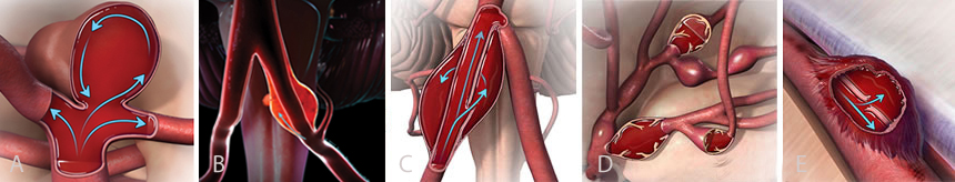
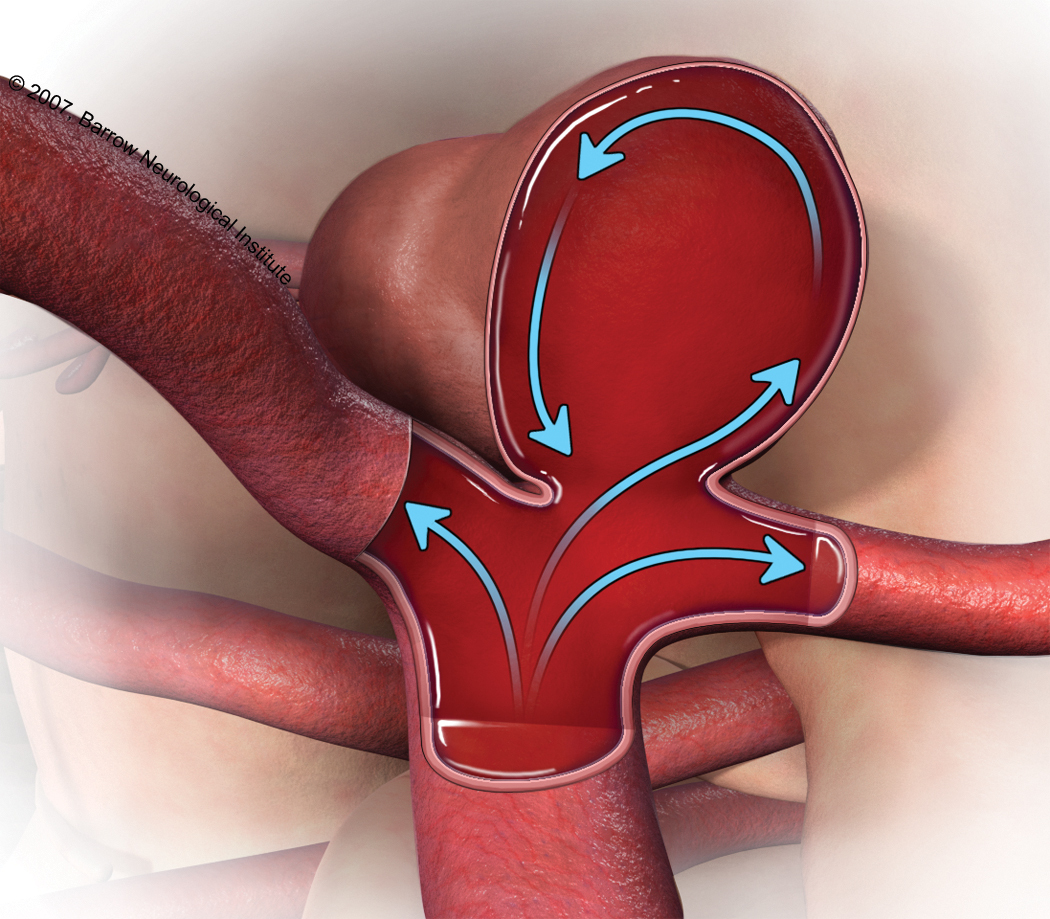
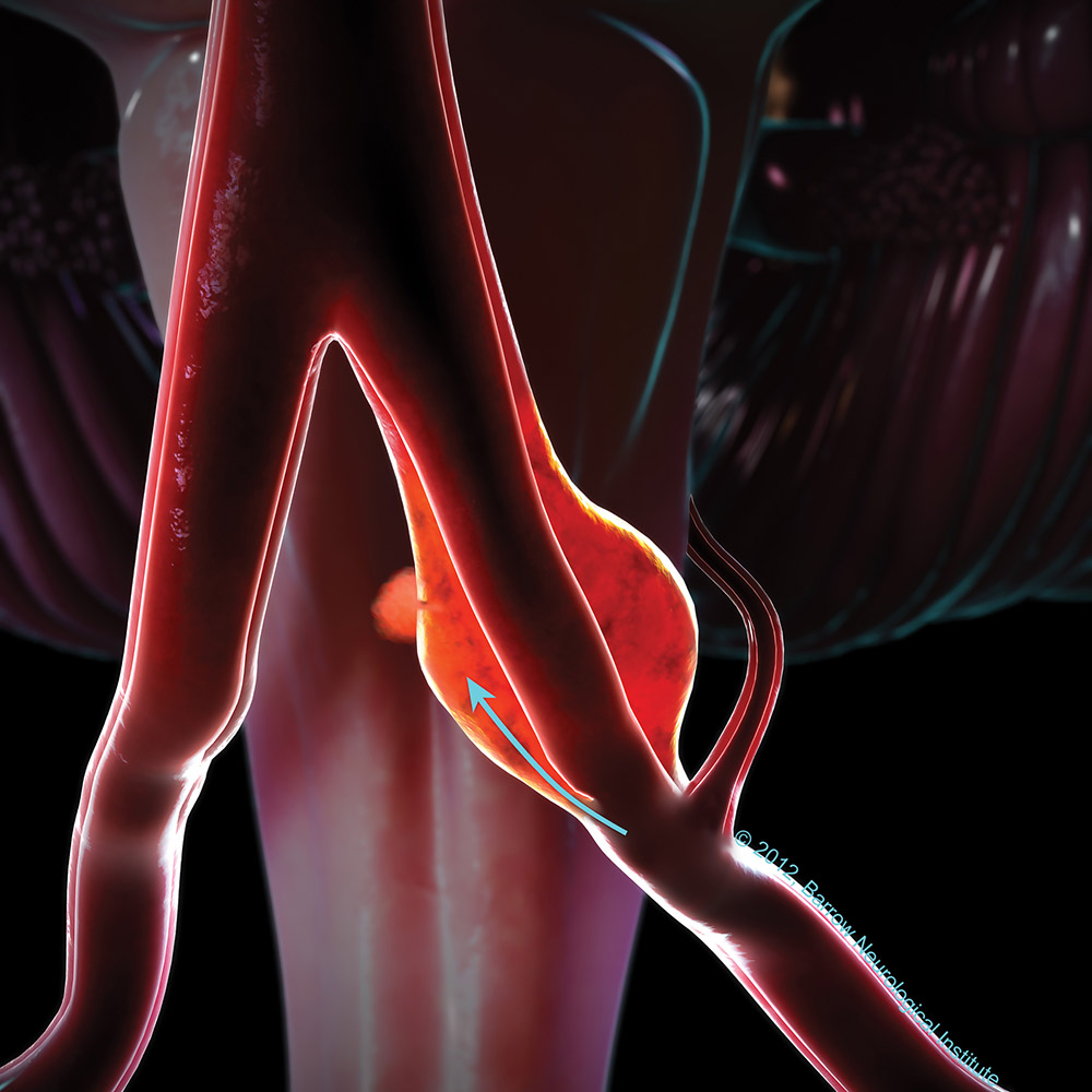
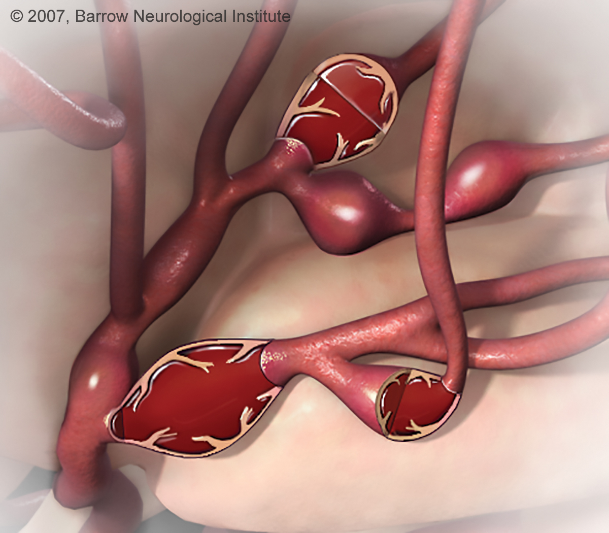
As for Imaging vs angiograms, angiograms allow the specialist to see better than the imaging provides. I had over a dozen CTs whilst in ICU, BH says it’s closer to two dozen. Either count, my Neurosurgeon stays away from them for me and uses the MRAs. When I went for my psych testing after rupture because my brain was “black” ( no thought processing) the dear Psychologist said I could light up the county with all the CTs I had, my Neurosurgeon agreed.

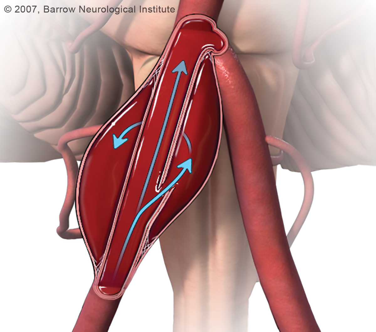
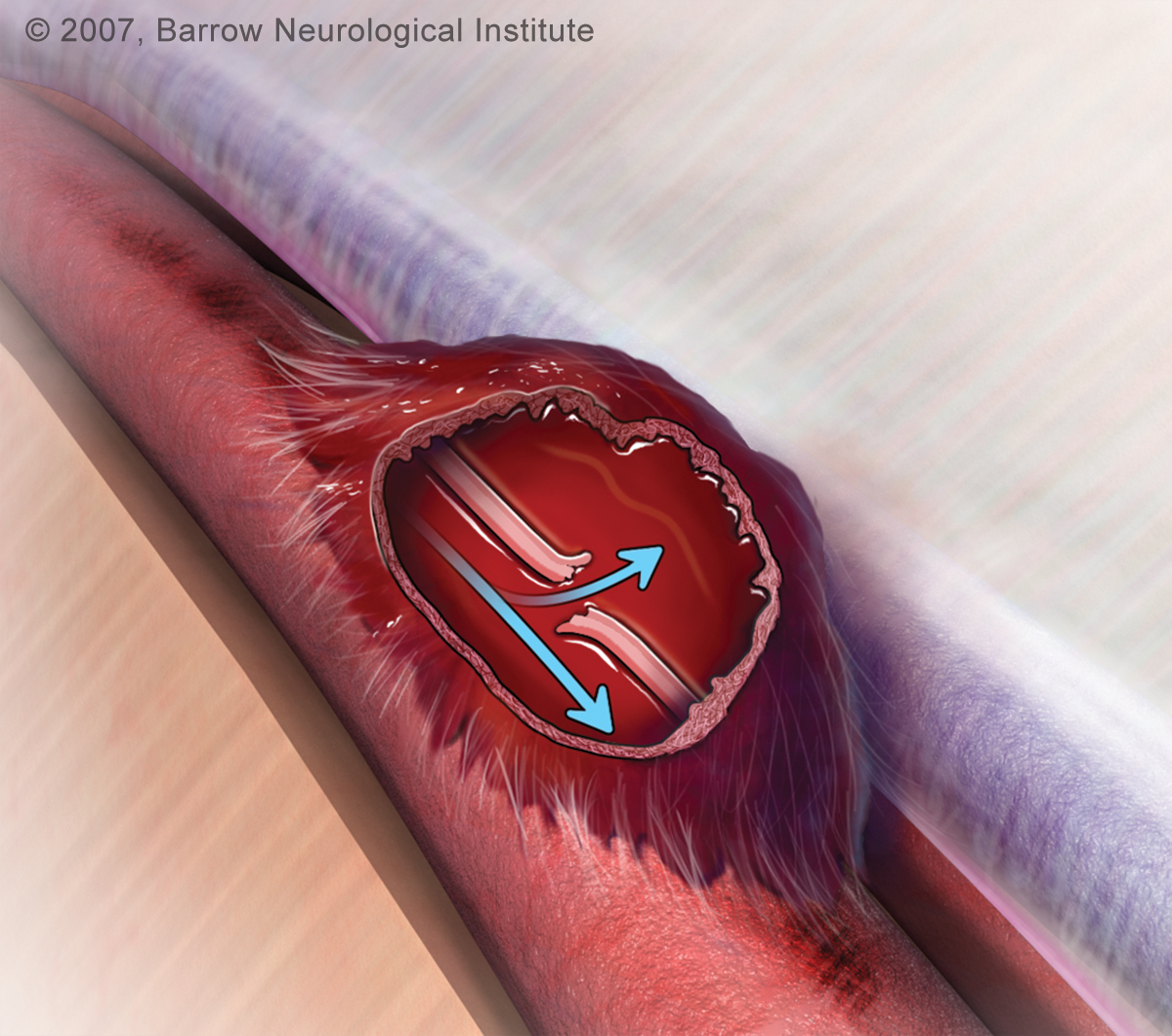
Will your surgeon do a follow up angiogram? I can’t remember if they do one for craniotomy procedures or just use imaging. Obliteration as I understand it is the lack of blood flowing into the aneurysm. The aneurysm is always with us, it just stops being fed by the device or procedure done to stabilize it. If the neck or a portion of it is still being fed, but the device hides that little portion it won’t be seen. The way I understand it is the images are to see not only the aneurysm but if the device be it stents, clips, or coils or all is where it’s supposed to be. I may be way off base with this thinking and will definitely ask my surgeon in June. I wonder if your surgeon feels a CT can get around the artifact? Maybe ask the tech that does your imaging, I’d really like to know. I’ve had images where it states the aneurysm is obliterated, yet I still have to get the little bugger fixed. Maybe I don’t understand the definition of obliterated the way the docs understand it…

I think Merl @ModSupport could probably answer your question much better than I am.

@Tony_P you are right! It really looks like a happy face, that’s great! Good for you!

All the best,
Moltroub

1 Like

I’m not sure how I missed this the other day. That is an impressive scar! I haven’t had brain surgery, but lots of other surgeries. I look at them as telling the story of our lives, whether from surgery or accidents. That scar tells me that you’re pretty bad*ss! I’ll let the brain surgery experts talk about the specifics of recovering from that, which definitely has different issues.

Sharon from ModSupport

2 Likes

Hmmm, not real sure about that one Moltroub
BUT
I had an MRI scan with a reported ‘artefact’, it was then recommended I have a CT to confirm all was OK behind what the artefact was hiding. I was having symptoms the question was ‘Is the artefact hiding the cause?’ In my case it wasn’t hiding anything, but they needed to check, hence the CT scan.

Also angios show what is going on within the vessel being examined, but what about other vessels within the brain? A CT (Often with contrast dye) will show up any other vessels/abnormalities within the entire brain and not solely that vessel.

Merl from the Modsupport Team

1 Like

Thanks Merl! For me the MRA does the brain vessel check, only the pituitary adenoma has to be special order.

Best, Moltroub

1 Like

All of my CTA/MRA for the past 5 years have shown no residual flow into the aneurysm and then I have an angiogram done and there the regrowth is. Twice as large as the clipped portion. Bendok told me that the clip is throwing off so much artifact on CTA and MRA that radiology cannot see directly below the clip, so the residual/regrowth was missed. Angiography obviously visualizes everything. I just don’t know what they think they will see on CTA now? The clip is still there throwing off artifact…

I will have an angiogram at 1 year, and I will not be surprised if the findings are different from today’s CTA.

@phoenix33 a year is a good bit of time to wait isn’t it? My routine after rupture went something like MRA, Angiogram, Repair, MRA, angiogram; repeat. This last time the MRA told her all she needed to know and we skipped the angiogram and went straight to repair. Each image has had notation of artifact being caused by something. The “something” is never exactly the same LOL. I’ve never spoken to a radiologist about artifacts, and I can’t remember if I asked my Neurosurgeon but I think it shows up as a blur, shadow or hazy area in the area we would most certainly like them to be able to see.

Once again @phoenix33, you have given me food for thought and another question added to the list! You are priceless to me!

All the best and thank you so very much, again!
Moltroub

1 Like

Hi @Moltroub! Well, as predicted, not much seen on CTA. The flow diverter has implanted nicely, and is wide open with excellent flow. Unfortunately, we cannot see the aneurysm itself due to the clip. Bendok did say if we did an angiogram today he feels the aneurysm would be obliterated. Ha! Exactly the words of the great Robert Spetzler. Only time will tell…1 year angiogram will be here altogether too soon. He did say that I made the right decision because this residual was “going to harm me,” but we kind of knew that going in. I believe it was just his way of reinforcing that treatment vs observation was the best option.

@phoenix33, I’m glad the stent is doing it job best they can tell, that’s good news! My Neurosurgeon has never told me my aneurysm is obliterated this last time she just was very excited and said she thinks she got it fixed. I wonder why he is waiting for a year and not six months, so you know? I’m guessing he is confident… Do you have a set of rules he has advised for the year?

By the way, I’m really glad you’re sharing your journey with us. As you have taught me blister aneurysms are very rare, I think you even pointed out only one other member has had one. When one looks up cerebral aneurysms, it’s always saccular, fusiform or mycotic. I’ve never read a paper that goes into all the subsets of those three groups. If anyone has ran across one, I’d sure like to read it!

Hugs,
Moltroub

1 Like

Types of Cerebral Aneurysms

A true aneurysm is an expansion of a blood vessel wall involving all layers of the wall. The two most recognized types of true aneurysms are saccular and fusiform, although mycotic, pseudo, and blister represent the rarer types of aneurysms. Most aneurysms occur sporadically, however they can be associated with other medical conditions such as Marfan’s syndrome, fibromuscular dysplasia (FMD), polycystic kidney disease, and Ehlers-Danlos syndrome. In rare instances cerebral aneurysms are a heritable condition that runs in families. Current guidelines recommend screening tests for cerebral aneurysms if two or more first-degree relatives in one family are known to have harbored cerebral aneurysms.

Joe Niekro Foundation - Four Types of Celebral Aneurysms
Types of aneurysms (A) saccular, (B) dissecting, (C) mycotic, (D) pseudo and (E) blister

Saccular

Joe Niekro Foundation - Types of Celebral Aneurysms

The “saccular” or “berry” aneurysm is the most common type of aneurysm, and it’s the one we refer to when we think of “brain aneurysms” in general. Berry aneurysms are ones that look like sacs or berries sticking out of a side of a blood vessel wall. Most of these have a “neck” region, although sometimes a neck cannot be readily defined in this type of aneurysm, even at the time of surgery. Berry aneurysms are associated with growth and rupture. They are also the most common cause of non-traumatic subarachnoid hemorrhage (SAH), and are attached to the larger portion (dome). Saccular aneurysms develop along weak spots in the arterial wall.

FUSIFORM

Joe Niekro Foundation - Types of Celebral Aneurysms

The “fusiform” (dissecting) aneurysm, is less common than the saccular aneurysm and looks like the blood vessel is expanded in all directions. Dissecting aneurysms form from injuries to the innermost layers of the blood vessel, such as after a traumatic injury or from the formation of atherosclerotic (fatty) plaque. Fusiform aneurysms don’t have a “neck” region, and they seldom rupture. They rarely present with SAH but can cause strokes and, when they grow in size, can become symptomatic.

MYCOTIC

Joe Niekro Foundation - Types of Celebral Aneurysms

The “mycotic” or “infectious” aneurysm is very rare and is a saccular aneurysm that arises from an artery that has had a certain part of the wall affected by a source of infection usually originating from somewhere else in the body (e.g., the heart) and spreading to the brain blood vessel by the blood stream. Mycotic aneurysms are caused by infectious agents, often in association with subacute bacterial endocarditis. Multiple mycotic aneurysms, found along the distal (superficial) portions of the brain arteries, are common.

PSEUDO (DISSECTING)

Joe Niekro Foundation - Types of Celebral Aneurysms

A false or “pseudo-aneurysm” brain aneurysm is an expansion of a blood vessel wall that does not involve all layers of the wall. Most commonly, it involves the outermost layers of the brain artery only, and usually follows injury or tearing of the vessel wall (referred to as a “dissection” or “laceration”). Pseudoaneurysms usually form in the regions where the falx or tentorium is near cerebral arteries. Ballooning may occur on one side of the artery wall, or it may block off or obstruct blood flow through the artery. Dissecting aneurysms usually occur from traumatic injury but can also form spontaneously. Treatment is determined by the shape and location of the aneurysm.

BLISTER

Joe Niekro Foundation - Types of Cerebral Aneurysms

Blister aneurysms can occur anywhere on the brain blood vessels but most commonly form on the internal carotid artery and have a blister-like appearance on the vessel. These aneurysms may have an increased incidence of bleeding and higher mortality rate.

Your wish is my command… :wink:

@phoenix33 i wish you could see my smiling face as I say “Ok smarty britches, where’s the multi-lobe aneurysms?” And then I bust out laughing! Yes I say smarty britches, because I couldn’t think of the word “pants” and out came britches, it’s stuck.LOL.

All joking aside, this is really good information @phoenix33, thank you so much for posting it!

All the best
Moltroub

1 Like

It stops being funny when it starts being you…

I have to say I’ve never found another’s pain to be ‘funny’ but the man in question had to be shown via his own health situation how cutting his former comments had been. I know comprehending another’s pain can be near on impossible and I doubt any explanation of the level of pain would actually register with him. His attitude was ‘Well, just toughen up Princess’. Short of taking a ballpein hammer and hitting him with it (which I sure wish I had handy at the time :wink: ) he simply wouldn’t understand. But by using his own words, on his own present situation… …now that touched a nerve and he didn’t like that, not one little bit.

The man still has no clue what excruciating pain is like, none at all, but he’ll certainly think twice before making another ‘funny’ comment in my presence because I’ll have no issue in reminding him again.

Merl from the Modsupport Team